New breakthroughs in understanding the brain’s waste-clearing glymphatic system are opening doors to novel Alzheimer’s and Parkinson’s interventions, from lifestyle tweaks to future drugs.
The brain runs a nightly garbage haul that is now at the heart of Alzheimer’s and Parkinson’s research.
The brain is a greedy organ, burning about 20 percent of the body’s energy while weighing only three pounds. Yet for decades, scientists were baffled by a simple question: How does the brain get rid of its trash? Most tissues use lymphatic vessels to drain waste, but the brain appeared to lack them. The discovery of the glymphatic system in 2012 answered that question and rewrote the rules of neurobiology. Named for its dependence on glial cells and its similarity to the lymphatic system, this brain-wide network of perivascular channels is now a central player in the development of Alzheimer’s disease, Parkinson’s disease, and other neurodegenerative conditions. As researchers continue to decode its intricate machinery, a host of novel therapies and lifestyle interventions are emerging to keep the brain’s plumbing in tip-top shape.
The Brain’s Nightly Rinse
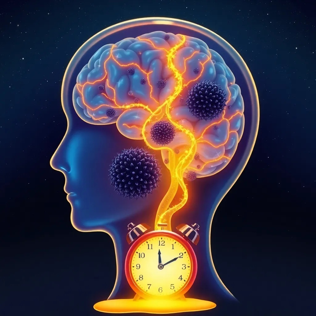
The glymphatic system is a sort of aqueduct for the mind. It begins where cerebrospinal fluid, the clear liquid that cushions the brain, flows through spaces around arteries. Specially organized water channels, aquaporin-4 or AQP4, carry that fluid across astrocytic endfeet into the brain’s dense interstitial spaces. Once inside, the CSF mixes with extracellular fluid, picks up metabolic waste such as amyloid-beta, tau, and alpha-synuclein, and then vessels along veins direct the contaminated fluid toward the meningeal lymphatic vessels for disposal. The entire loop operates in a constant cycle, but with a peculiar twist: it is far more active during sleep. In a landmark 2013 study published in Science, Lulu Xie and her colleagues at the University of Rochester reported that when mice slept, their interstitial space expanded by 60 percent. This increased cross-sectional area allowed cerebrospinal fluid to flow more easily and wash away amyloid-beta at roughly 1.4 times the rate seen in awake mice. The findings were a revelation. It offered a mechanistic explanation for why sleep deprivation is linked to Alzheimer’s dementia, and it highlighted sleep as a critical, non-negotiable neuroprotective behaviour.
Since then, researchers have refined the model. The glymphatic system is not a static tube, but a dynamic, pressure-driven network. Arterial pulsations play a key role, and slow waves during deep sleep create electrical oscillations that generate the cerebrospinal fluid flux. A 2022 paper in Nature Neuroscience, from the lab of Yunjuan Sun at the National Institute of Neurological Disorders and Stroke, showed that breathing rhythms modulate glymphatic flow at the skull base, further emphasizing that every respiratory cycle helps pump fluid through the brain. This finding has inspired new approaches, including voluntary nasal breathing techniques and home-based devices to enhance cerebrospinal fluid movement.
When the Binding Breaks
The link between glymphatic dysfunction and neurodegeneration is now one of the most actively researched areas in neuroscience. In Alzheimer’s disease, the accumulation of amyloid-beta and tau follows an almost predictable trajectory. Amyloid-beta clumps begin to form years before the first memory complaint, and tau tangles appear later, closer to symptom onset. Studies in mice and humans have consistently shown that glymphatic impairments worsen these pathologies. For example, a 2017 study led by Maiken Nedergaard and Jeffrey Iliff at the University of Rochester found that knocking out AQP4 in mice reduced solute clearance by roughly 70 percent and led to an age-related increase in amyloid burden. The same team later demonstrated that aged mice show a dramatic loss of AQP4 polarization, meaning the channels are unevenly distributed across astrocytic endfeet, causing fluid to leak into wrong parts of the brain. This age-dependent mislocalization is now considered a key driver in the sporadic form of Alzheimer’s, which accounts for over 95 percent of all cases.
Parkinson’s disease also bears the footprints of a failing cleanup system. Alpha-synuclein, a protein that misfolds and aggregates into Lewy bodies, is normally cleared via the glymphatic pathway. A study from the University of Oslo in 2021 used contrast-enhanced MRI to measure glymphatic function in patients with early Parkinson’s and identified a significant reduction in fluid clearance, even before the onset of severe motor symptoms. Similarly, research on traumatic brain injury has revealed that concussions can impair glymphatic flow for weeks. Repeated head impacts in animal models lead to sustained AQP4 downregulation and perivascular space collapse, accelerating tau aggregation. This has major implications for athletes and military personnel who experience repeated blows to the head. A 2020 article in Brain reported that retired football players with a history of concussions had enlarged perivascular spaces, indicating chronic glymphatic impairment. These findings are not just academic; they provide a potential target for early diagnosis and preventive treatment among at-risk groups.
Restoring the Flow: Emerging Therapies
The next wave of Alzheimer’s and Parkinson’s therapies may not try to clean out existing plaques, but rather fix the brain’s ability to cleanse itself. This approach is gaining traction among pharmaceutical developers and academic labs alike. At the highest level, lifestyle interventions, particularly sleep, remain the most effective way to preserve glymphatic function. But even the position of your head while sleeping matters. Researchers have reported that sleeping on the side, the lateral position, facilitates glymphatic clearance more effectively than sleeping on the back or stomach. Exercise is also potent. Aerobic exercise increases the amplitude of arterial pulsations and has been shown to enhance glymphatic influx in mice, according to a 2019 study in the Journal of Cerebral Blood Flow and Metabolism.
Now, pharmacological interventions. Several experimental drugs aim to bolster glymphatic function by targeting AQP4. Small-molecule activators that enhance the expression or polarization of AQP4 have been developed and shown to reduce tau pathology in mice. One such compound, identified by researchers at the University of Copenhagen and announced in 2023, was able to restore cognitive performance in a mouse model of tauopathy. Human trials are being planned. Also, intermittent CO2 exposure is generating buzz. Inhaling air containing 5 percent carbon dioxide for short periods causes vasodilation of the cerebral vasculature, increasing blood flow and pulsatility. A 2021 study in animals found that CO2 inhalation doubled the influx of CSF into the mouse brain, and the effect persisted for at least 30 minutes after exposure. The technique has not yet been tested in humans, but it offers a simple, low-cost approach that could be combined with other interventions.
Diagnostics also benefit. Contrast-enhanced MRI is now enabling clinicians to visualize perivascular spaces and measure glymphatic activity. Researchers at Thomas Jefferson University have developed an AI tool that automatically quantifies the dilation of perivascular spaces in T2-weighted images. In a 2024 proof-of-concept study, they demonstrated that this index correlates with cognitive decline and can predict progression to Alzheimer’s in patients with mild cognitive impairment with over 80 percent accuracy. Such biomarkers are crucial for selecting patients for future glymphatic therapies and for monitoring their response.
What are the challenges? The brain lacks a true lymphatic system in the classic sense, and researchers still debate the fluid dynamics. Some argue that CSF flow is driven by arterial pulsatility rather than AQP4. Others worry that CO2 exposure could cause vasodilation and raise intracranial pressure. The translational gap from rodents to humans is huge. Also, there is the ethical issue of who should be treated. If we can predict glymphatic decline at age 50, should we recommend daily CO2 inhalation? What about unapproved supplements claiming to boost the glymphatic system? The beauty and wellness industry is already jumping on the ‘brain drain’ bandwagon, with products such as ‘neuro-cleanse’ teas and head massagers claiming to enhance cerebrospinal fluid flow. Dermatological interest is also emerging, linking skin and brain clearance through the same lymphatic pathways.
The current emphasis on the glymphatic system is best understood as part of a long story of paradigm shifts in Alzheimer’s research. For more than two decades, the β-amyloid hypothesis held the field in an iron grip. Huge sums were invested in therapeutic antibodies that were designed to bind and remove amyloid plaques. But after a string of high-profile failures, including bapineuzumab, solanezumab, and crenezumab, the scientific community began to question whether plaque removal alone could rescue cognition. The controversial FDA approval of aducanumab in 2021, based on surrogates rather than definitive cognitive outcomes, and then lecanemab in 2023, which did show a modest 27 percent slowing of cognitive decline at 18 months, brought a complicated victory. Both drugs carry significant risks of brain swelling and microhemorrhages. Ironically, these results suggest that simply attacking plaques is insufficient. The brain’s clearance capacity, determined by the glymphatic system, may be an equally critical part of the equation. In that sense, glymphatic research is returning to a broader biological view of the brain, respecting its homeostatic cycles rather than treating it as a test tube containing a single misfolded protein.
The growing popularity of ‘brain detox’ and ‘sleep cleanup’ products in the wellness industry reflects how the glymphatic concept is rapidly seeping into consumer culture. Brands are selling neck pillows that claim to align perivascular spaces, sleep gummies infused with omega-3 and caffeine-free botanicals, and even CO2 ‘cleanse’ sessions. This pattern is similar to the hyaluronic acid boom of the early 2010s, when the molecule was touted as the ultimate anti-aging ingredient after initial veterinary studies. The product cycle tends to overhype before the evidence catches up. The most evidence-based glymphatic interventions remain as unglamorous as they come: regular deep sleep, consistent aerobic exercise, and avoiding alcohol and antihistamines that disturb sleep architecture. The true test of this field will come from large, long-term trials measuring hard outcomes like dementia incidence. Until then, the glymphatic system is an exciting biological target, but not a miracle cure. It is a reminder that the brain, like any organ, survives only if its waste is efficiently removed. And maybe that metaphor extends beyond our gray matter: our society, too, needs to clear away old dogmas to make space for new, evidence-driven approaches.